Gray Medical
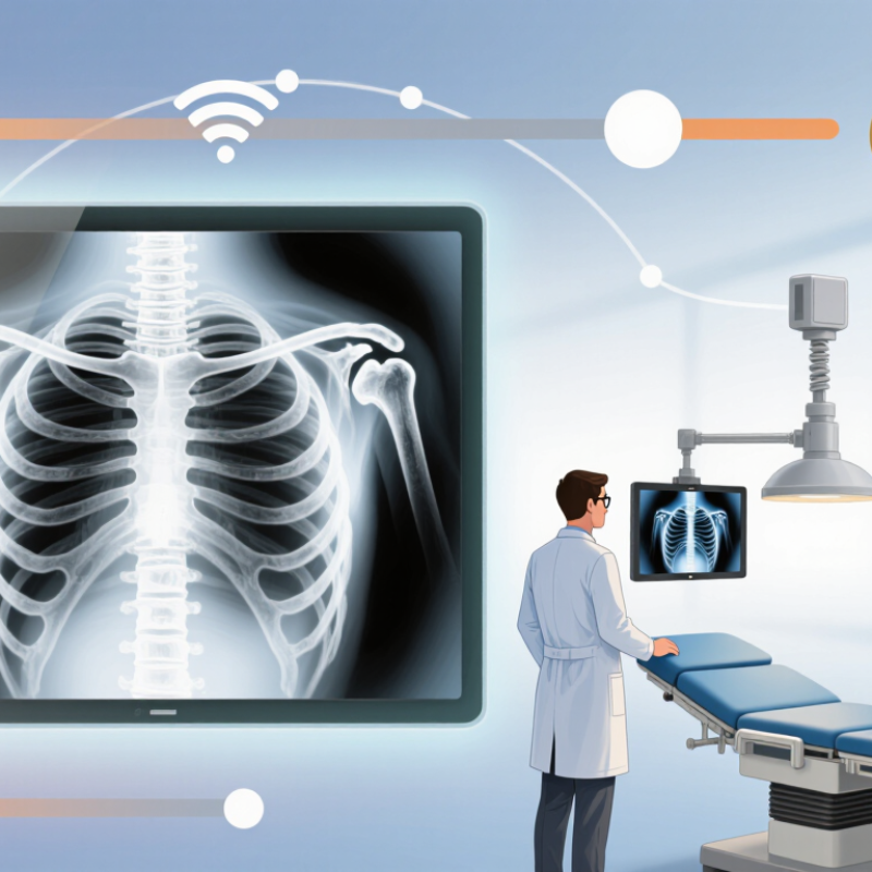
Why is digital imaging replacing traditional X-ray film? This question reflects a major change in everyday radiography. In many clinics, a digital detector captures an image within seconds. A radiographer can review it on a monitor beside the examination table. This shortens waiting times and helps clinicians respond sooner when urgent findings appear. Digital images also travel through secure picture archiving and communication systems. A specialist can assess them remotely without shipping a physical envelope. That practical difference matters in busy hospitals and smaller community facilities.
Digital systems offer more than speed. Their software can adjust brightness, contrast, and magnification after exposure. These tools may clarify a subtle fracture or improve visibility around dense anatomy. In my experience? That phrase requires caution, because results depend on positioning, detector quality, and staff training. Digital radiography can reduce repeat examinations when exposure settings are appropriate. However, automatic processing may hide technical errors rather than correct them. Good image quality still begins with careful positioning and radiation protection.
Film remains useful in some settings, especially where budgets, electricity, or technical support are limited. Digital equipment requires investment, maintenance, data protection, and reliable network access. It can also encourage unnecessary imaging if convenience replaces clinical judgment. Therefore, replacement is not simply a technological victory. It is a balance between diagnostic value, patient safety, workflow, and long-term cost. This article examines the evidence, practical experience, and limitations behind digital imaging’s growing role, while acknowledging that no system is perfect.
Traditional X-ray film is a photographic recording medium made from silver halide crystals. These crystals sit inside a light-sensitive emulsion on a polyester base. During an examination, X-rays pass through the patient and reach the film cassette.
Most cassettes use intensifying screens. The screens convert X-ray energy into visible light, which exposes the film more efficiently. A latent image forms, but it cannot be seen immediately. The film must enter a darkroom, where developer reveals the image, fixer stabilizes it, and washing removes residual chemicals. Drying follows. A darkroom shaped the image.
The World Health Organization estimates that about 3.6 billion diagnostic X-ray examinations occur globally each year. That scale explains why film workflow matters. Film can provide strong contrast and a permanent physical record, but exposure errors are difficult to correct. A slightly overexposed area may lose useful detail forever. The International Atomic Energy Agency notes that imaging quality and radiation protection must be managed together, not separately. In practice, film handling also creates delays, chemical waste, storage demands, and repeated work when images are misplaced. The process was not elegant. Yet it trained generations of radiographers to read density, positioning, and artifacts carefully. That discipline remains valuable, although relying on memory alone can invite mistakes.
Digital imaging begins when X-rays pass through the patient and reach a digital detector. Inside the detector, a scintillating layer changes X-ray energy into visible light. Photodiodes then convert that light into electrical signals. Each pixel records a different signal according to the tissues it represents. A computer converts these measurements into shades of gray. Within seconds, the image appears on a clinical workstation. Small details matter. An angled detector or patient movement can blur anatomy and reduce diagnostic value. The rapid preview helps radiographers check positioning before the patient leaves the room.
Image processing then adjusts brightness, contrast, and edge visibility for clearer inspection. It can highlight bone structure while preserving differences in soft tissue density. The system also stores exposure information and transfers images through secure clinical networks. Digital images are not automatically better. Excessive processing may create artificial edges or hide subtle findings. A bright, sharp-looking image can still result from poor positioning or unsuitable exposure. Experienced radiographers review image quality, while qualified clinicians interpret the anatomy and clinical context. Dose monitoring remains important because easy image repetition may encourage unnecessary exposures. In practice, reliable imaging depends on careful positioning, tested equipment, appropriate settings, and human judgment.
Digital imaging changes the examination room in practical ways. Images appear within seconds, not after chemical processing and drying. It is immediate. A radiographer can check positioning before the patient leaves. That reduces avoidable return visits and supports faster clinical decisions.
The World Health Organization and IAEA estimate that about 3.6 billion diagnostic X-ray examinations occur worldwide each year. At this scale, workflow efficiency matters. Digital files move securely between imaging rooms and authorized clinicians. Film requires physical storage, transport, and manual retrieval. NHS England recorded more than 46 million diagnostic imaging tests in 2023–24, showing why accessible records can ease pressure on busy services.
Digital detectors also offer broad exposure latitude and software-based contrast adjustment. A radiologist can inspect bone edges, soft-tissue regions, and small fractures without exposing the patient again. The IAEA’s radiation-protection guidance stresses optimization, however; digital systems do not automatically lower dose. Overexposure can become less visible, because the image may still look acceptable. This is an important weakness.
There is less chemical waste, too. Film processing uses developer, fixer, water, and darkroom equipment. Digital workflows remove most of those materials. Yet electronic systems need calibration, cybersecurity controls, backups, and trained staff. In practice, poor positioning still produces poor images. Technology cannot repair every human mistake.
Why Is Digital Imaging Replacing Traditional X Ray Film?
Digital imaging offers faster results, easier storage, and simpler image sharing. Yet its convenience can hide serious technical challenges. A digital detector may produce a clear image even when the patient receives unnecessary radiation. This is known as dose creep. It can develop quietly when staff adjust exposure settings for brighter images.
Infrastructure is another limitation. A busy clinic needs stable electricity, reliable networks, calibrated monitors, and secure data storage. A failed detector can interrupt examinations for hours. Poor display calibration may also hide small fractures or soft-tissue details. Digital images are not automatically accurate. They still require trained radiographers and qualified clinicians.
Cybersecurity and privacy create additional responsibilities. Patient images contain sensitive medical information, so access must be controlled and monitored. Different systems may not communicate smoothly, causing delays or duplicate records. Training is often underestimated. New staff may understand the software but miss positioning errors or motion artifacts. That matters.
Traditional film had weaknesses, but it made certain mistakes visible. Digital systems can make errors look polished. Clinical teams should review exposure indicators, repeat-image rates, and equipment performance regularly. Even experienced professionals can trust automation too much. More reflection is needed before assuming that faster imaging always means better care.
Digital imaging is replacing traditional X-ray film because it changes more than image storage. A radiograph can appear within seconds, allowing clinicians to assess urgent injuries faster. Images can also move securely between departments, hospitals, and remote specialists. This supports earlier consultation, especially where radiologists are limited. The WHO’s Global Initiative on Radiation Safety in Health Care Settings reports about 3.6 billion diagnostic X-ray examinations worldwide each year. At this scale, faster access can influence many treatment decisions.
Digital tools also improve comparison and continuity of care. A clinician can place today’s image beside an older scan and examine subtle changes. Electronic records reduce physical storage and avoid film-processing chemicals. Yet digital does not automatically mean safer care. The UNSCEAR 2020/2021 Report indicates that CT represents roughly 10% of diagnostic examinations but contributes about 60% of collective radiation dose. Better screens cannot replace careful justification, dose optimisation, and trained interpretation. I still see one blind spot: convenient imaging may encourage unnecessary testing.
Tips: Ask what clinical question the scan should answer. Confirm that previous images are available. Request a dose explanation when CT is recommended. For healthcare teams, audit repeat scans, monitor reporting delays, and maintain clear referral protocols. Small workflow failures remain important.
Digital imaging enables immediate image review, electronic storage, rapid sharing, and dose optimization across healthcare settings. The chart shows typical effective radiation doses for common digital imaging examinations. Actual dose varies with patient size, equipment, and imaging protocol; digital imaging does not eliminate radiation exposure, but it supports more efficient and better-controlled care.
Reference basis: published patient-dose estimates from professional radiology guidance and clinical imaging reference ranges.
Images appear within seconds, without chemical processing, drying, or darkroom handling. It is immediate. Clinicians can check positioning before the patient leaves.
Digital files can move securely to authorized clinicians. This reduces physical storage, transport, and manual record retrieval. Faster access may support quicker decisions.
No. Digital systems can tolerate a wider exposure range. An image may look acceptable after unnecessary radiation. This problem is called dose creep.
Dose creep happens when exposure settings gradually increase. Staff may seek brighter or clearer images. The change can remain unnoticed without regular monitoring.
It can reduce some repeats by showing positioning immediately. Poor positioning, movement, or technical mistakes still cause retakes. Technology cannot fix every human error.
It needs stable electricity, reliable networks, calibrated monitors, secure storage, and maintained detectors. A detector failure can stop examinations for hours.
Digital images contain sensitive medical information. Access should be restricted, monitored, backed up, and protected through strong cybersecurity controls. Convenience creates responsibility.
No. Poor monitor calibration can hide small fractures or soft-tissue details. Trained staff must assess positioning, exposure indicators, and motion artifacts.
It removes most developer, fixer, water, and darkroom waste. However, electronic systems still consume energy and require equipment maintenance. The benefit is real, but incomplete.
It is faster and easier to share, but not automatically safer or more accurate. Clinical teams should review repeat rates and equipment performance regularly. Faster care can still be flawed.
Why is digital imaging replacing traditional X-ray film? Traditional X-ray film records radiation on a physical sheet that must be chemically processed, stored, and manually reviewed. Digital imaging, by contrast, uses electronic detectors to capture X-ray data and convert it into computer-readable images. These images can be displayed quickly, adjusted for clearer viewing, and shared securely with authorized medical professionals.
Digital X-rays offer faster results, easier storage, reduced processing materials, and the possibility of lowering radiation exposure through improved image efficiency. They can support more convenient diagnosis, communication, and long-term patient care. However, the technology also requires costly equipment, technical training, regular maintenance, and strong safeguards for digital patient information. Image quality may also depend on detector performance and proper operating procedures. Despite these challenges, digital imaging is transforming medical diagnosis by making X-ray services faster, more flexible, and better integrated into modern healthcare systems.